
The Missing Link Between Glyphosate and Human Health
How Bifidobacteria Continue the Chemistry That Plants Begin

Introduction
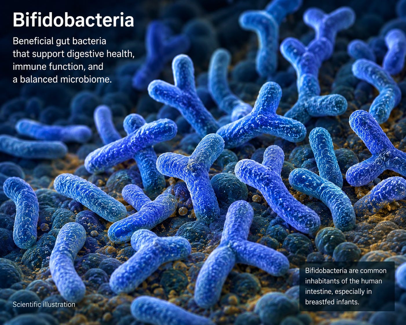
The last article ended with a question hiding inside a fact.
Glyphosate works by blocking the shikimate pathway — the biochemical route that plants and bacteria use to build aromatic amino acids. Humans don’t have this pathway, which is why the herbicide was declared safe for us. We don’t make these molecules. We get them from food.
That’s the official story but it isn’t the whole story.
Phenylalanine, tyrosine, and tryptophan are not inert nutrients that the body files away after a meal. They are starting materials, building blocks. The chemistry that begins in the shikimate pathway of a plant or a soil bacterium continues inside the human body — and inside the human microbiome with our microbial partners in life. These three molecules become dopamine, serotonin, melatonin, folate, and dozens of other compounds that regulate how we think, sleep, fight infection, and develop from birth.
The shikimate pathway is critical to life because those three amino acids are the entry point to an enormous biological network of molecules. And much of that network runs through the gut.
This article follows the chemistry forward.
The Second Half of the Shikimate Pathway
The shikimate pathway doesn’t end with just three molecules. What happens next is rarely discussed. One of the observations that got me interested in this is the first place was the observation that my husband John had some genetic snips involving phenylalanine and metabolism and symptoms in the first four years of his life that were very similar to what is described in a genetic disease called PKU. With this inborn error of metabolism children are put on a diet that eliminates phenylalanine entirely.
Phenylalanine, tyrosine, and tryptophan are classified as essential amino acids, which means the human body cannot synthesize them. We depend entirely on plants, bacteria, and the food we eat to supply them. But essential understates what they are. These are not building blocks waiting to be assembled into protein. They are the starting point for an entire tier of human biochemistry.
Phenylalanine converts to tyrosine. Tyrosine converts to dopamine, then to norepinephrine and epinephrine — the catecholamines that drive alertness, stress response, and the basic mechanics of motivation. Tyrosine is also the raw material for melanin, and for something less often mentioned in this context: the thyroid hormones T3 and T4. The thyroid takes tyrosine, attaches iodine to it, and produces the hormones that regulate metabolism, body temperature, heart rate, and fetal brain development. One amino acid. Two major hormonal systems. Both entirely dependent on a pathway that glyphosate blocks.
Tryptophan follows a different route. Most people know it as the precursor to serotonin. But serotonin is not the end of that road. Serotonin converts to melatonin, which governs sleep and circadian rhythm. Tryptophan also feeds the kynurenine pathway, which produces signaling molecules involved in immune regulation and inflammation. And a significant portion of tryptophan metabolism happens not in human cells but in the gut — processed by bacteria into indoles, a class of compounds that communicate directly with the intestinal lining, the immune system, and the brain.
Folate belongs in this company as well, though its origin is less familiar. The shikimate pathway produces chorismate, which bacteria use to synthesize para-aminobenzoic acid, or PABA — the structural core of folate. Folate is not just a B vitamin you get from leafy greens. It is a molecule that certain gut bacteria manufacture from shikimate-derived precursors. It sits at the center of one-carbon metabolism, the biochemical process that drives DNA synthesis, DNA methylation, and neurotransmitter production. We will come back to folate.
The full list of what these three amino acids become reads like a catalog of human regulatory biology: dopamine, norepinephrine, epinephrine, serotonin, melatonin, T3, T4, melanin, indoles, folate, polyphenol metabolites, quinones, immune signaling molecules. These are not minor compounds. They are the molecules that determine how the brain develops, how the immune system responds, how the body maintains itself across a lifetime.
And many of the transformations that produce them do not happen in human tissue. They happen in the gut.
Meet the Bifidobacteria
Not all gut bacteria are the same. This seems obvious, but the implications are routinely underestimated. Different species occupy different ecological niches, eat different substrates, produce different metabolites, and matter at different stages of life. Lumping them together as “good bacteria” is like saying all farmers grow food — technically true, and nearly useless as information.
The genus Bifidobacterium contains over fifty species. Five of them show up repeatedly in the research on human health, and each one tells a different part of the story.
Bifidobacterium infantis is the first. It colonizes the infant gut and is exquisitely adapted to human milk — specifically to the human milk oligosaccharides, the complex sugars that a nursing infant cannot digest but B. infantis can. It is not an accident that human milk contains sugars a baby cannot use. Those sugars are food for B. infantis, and B. infantis, in return, produces acetate and other short-chain fatty acids that fuel intestinal cells, lower gut pH to exclude pathogens, and begin the long process of training the infant immune system. This is a designed partnership. We will examine it in more detail shortly.
Bifidobacterium bifidum shares the early-life niche but brings different tools. It produces enzymes that break down mucins — the glycoproteins that form the intestinal mucus layer — which allows it to occupy the mucosal interface directly. Location, location, location. A bacterium living at the mucus layer is living at the border between the microbial world and human tissue. B. bifidum at that interface means a trained, tolerant immune response at exactly the right location.
Bifidobacterium breve is versatile across life stages and notable for its role in polyphenol metabolism. Plant polyphenols — the compounds that give berries, green tea, and dark chocolate their biological activity — are largely inaccessible to human enzymes. B. breve and related species transform them into bioavailable metabolites. The antioxidant activity attributed to polyphenol-rich foods depends substantially on this microbial conversion step.
Bifidobacterium longum is among the most extensively studied species in the genus and persists across the human lifespan, from infancy into old age. It is a generalist with notable capacity for tryptophan metabolism — producing indole derivatives that signal to the intestinal lining and influence the gut-brain axis. B. longum has also been studied in the context of stress and anxiety, with some of the more rigorous human trial data in the probiotic literature.
Bifidobacterium adolescentis colonizes primarily in adolescence and adulthood. It is an efficient producer of folate and acetate, and it is particularly active in fermenting resistant starches and plant fibers that other species cannot process. It also appears in the research for a reason relevant to this series: among the Bifidobacteria, B. adolescentis shows relative resistance to glyphosate compared to other members of the genus. It is my understanding from some of the research that it is the only Bifidobacteria that doesn’t produce plasmalogens, a special class of membrane phospholipids important to the brain and heart. Why that is, and what it means, is one of the questions this article will leave open.
Bifidobacteria as Microbial Chemists
The previous section introduced five species. This one sets the species aside and looks at what they collectively do — because the more useful frame is not which organism performs a reaction, but what chemistry gets performed, and what depends on it.
Bifidobacteria are not passive residents of the gut. They are active participants in human metabolism, running biochemical conversions that human enzymes cannot perform. Several of these conversions sit at the center of the biology this series has been building toward.
Aromatic amino acid metabolism
Bifidobacteria take phenylalanine, tyrosine, and tryptophan — the three molecules the shikimate pathway produces — and continue working on them. They deaminate, decarboxylate, and hydroxylate these substrates, producing intermediate compounds that feed downstream human pathways. This is the handoff point. The plant or soil bacterium produces the aromatic amino acid. The gut microbiome receives it and begins the next set of transformations. Human enzymes then take those intermediates and complete the synthesis of neurotransmitters, hormones, and signaling molecules.
Disrupt the middle step and the downstream products become scarce — even if dietary intake of the aromatic amino acids is adequate. Adequate intake of a precursor is not the same as adequate production of what the precursor becomes. It would be like having a car factory but the employees who build the engine are on strike or missing…then you can’t build the car.
Indole production
Tryptophan is used to synthesize serotonin in the enterochromaffin cells in the gut. But any unused tryptophan is available for microbial metabolism, and Bifidobacteria — particularly B. longum — convert it into indoles. This might sound insignificant because so much is used in serotonin but it’s not.
Indoles are important signaling molecules. They bind to the aryl hydrocarbon receptor (AHR), a transcription factor expressed throughout the gut, the liver, the lungs, and the brain. The AHR sits at the interface between the environment, the microbiome, diet and the immune system. When indoles activate this receptor in intestinal cells, the response includes tightening of the epithelial barrier, modulation of local immune activity, and production of IL-22, a cytokine involved in mucosal repair. Indoles produced in the gut also cross into systemic circulation and have been detected in the brain, where their signaling roles are still being characterized.
The gut lining communicates with the immune system partly through these molecules that Bifidobacteria make from an amino acid that plants provide through this critical shikimate pathway.
Folate (B9) synthesis
Bifidobacteria synthesize folate from scratch. The pathway runs through chorismate — a shikimate pathway product — to para-aminobenzoic acid, or PABA, the structural core of the folate molecule. From PABA, bacteria complete the synthesis of folate de novo — producing a vitamin inside the human colon, without any contribution from diet or supplementation.
How much of human folate status depends on this microbial production is not precisely known. The research has focused primarily on dietary folate and supplemental folic acid, with microbial production treated as a minor contributor. But honestly, that’s not really known. And that assumption deserves more scrutiny than it has received.
It also deserves scrutiny alongside a second disruption that has been running in the opposite direction. Since the 1990s, folic acid — the synthetic form of folate used in fortified foods — has been added to flour, cereals, and grain products across most of the industrialized world. Folic acid is not the same molecule as the folate that plants produce or that gut bacteria synthesize. It requires an extra enzymatic conversion step to become biologically active, and at high doses it can compete with and inhibit the very enzymes that process natural folates. The possibility that we have simultaneously disrupted microbial folate production through agricultural chemicals and masked that disruption with a synthetic substitute that creates its own metabolic interference has not received the attention it warrants. We will return to this in the folate section.
Polyphenol transformation
Plant polyphenols arrive in the colon largely intact because human digestive enzymes cannot break them down. Bifidobacteria — particularly B. breve — possess the enzymatic machinery to transform these compounds into smaller molecules that human cells can absorb and use. The anti-inflammatory and antioxidant effects associated with diets rich in berries, green tea, olive oil, and dark chocolate are substantially mediated by this microbial conversion step.
A polyphenol-rich diet without the bacteria to process it delivers less than the research on those foods would suggest. The bacteria are part of the mechanism.
Acetate and cross-feeding
Bifidobacteria are primary producers of acetate in the gut ecosystem. Acetate is a short-chain fatty acid, and in the gut it serves two functions simultaneously: it feeds the colonocytes — the cells lining the colon — and it is the substrate that other bacterial species, particularly butyrate producers, use to make butyrate. Butyrate is the preferred fuel of colonocytes and a potent regulator of intestinal inflammation and barrier integrity.
Bifidobacteria do not produce butyrate directly. They produce the acetate that makes butyrate production possible. Remove Bifidobacteria and you do not just lose their direct metabolic outputs — you destabilize the cross-feeding network that supports the organisms downstream of them.
Barrier signaling
The intestinal barrier is not a static wall. It is a dynamic interface maintained by constant signaling between the epithelial cells, the immune cells beneath them, and the microbial community above them. Bifidobacteria contribute to this maintenance through multiple routes: acetate signals directly to epithelial cells to strengthen tight junctions; indoles activate barrier-protective responses through the aryl hydrocarbon receptor; and the mucosal colonization of B. bifidum positions that species to influence barrier function at close range.
When the barrier fails — when tight junctions loosen and microbial products cross into systemic circulation — the downstream consequences include chronic immune activation, systemic inflammation, and disruption of the signaling environment in which neurotransmitter and hormone production occurs. Barrier integrity is not a separate topic from brain chemistry. They are connected, and Bifidobacteria sit at the junction.
Human Milk Reveals Nature’s Priorities
If you want to understand what biology considers important, look at what it protects most carefully.
Human milk is the most studied food in existence, and it contains something that puzzled researchers for decades. Alongside fat, protein, and lactose, human milk is rich in complex sugars called human milk oligosaccharides — HMOs. There are more than two hundred distinct HMO structures in human milk. They are the third most abundant solid component after fat and lactose. And a nursing infant cannot digest them.
This is not an accident or an evolutionary oversight. A molecule that costs the mother significant metabolic resources to produce, present in quantity in every feeding, that the infant cannot use — that molecule is not there for the infant. It is there for the microbiome.
Specifically, it is there for Bifidobacterium infantis.
B. infantis is uniquely equipped among gut bacteria to consume HMOs. It carries a cluster of genes — sometimes called the HMO utilization island — that encode the enzymes needed to import and break down these complex sugars completely, capturing the energy and releasing the component parts. Other bacteria can process HMOs partially. B. infantis processes them entirely, and it does so precision.
What B. infantis produces in return is not incidental. As it ferments HMOs, it releases acetate, which acidifies the gut environment and suppresses pathogen growth. It produces compounds that signal directly to intestinal epithelial cells to strengthen the barrier. It generates molecules that educate the developing immune system — teaching it, in the earliest weeks of life, the difference between commensal bacteria and threats. The infant immune system is not born knowing this. It learns it, and B. infantis is among its first teachers.
The relationship between human milk and B. infantis is one of the clearest examples in human biology of a co-evolved partnership. The mother produces a substrate she cannot use, to feed a bacterium her infant needs, to produce metabolites that protect and develop systems the infant cannot yet protect and develop on its own. We are more microbial than we are human and this is part of that initial development of our microbial selves. For years the ratio of microbial to human cells was cited as ten to one. More recent and more careful counting puts it closer to two microbial cells for every human one — and the ratio of microbial genes to human genes remains overwhelmingly microbial. We are not outnumbered by our microbial partners. We are intertwined with them, and we cannot function without them. In fact, in terms of genes and metabolic functions the microbiome provides 100-300 times more genes and functions that our own genome. Human milk is nature’s clearest acknowledgment of the relationship, the microbial community of the infant is essential to long term health.
The microbiome is not a bystander. Rodney Dietert, PhD has published on the Completed Human theory that essentially we are incomplete without our microbial partners.
This matters for what comes next. The partnership encoded in human milk establishes the Bifidobacteria-dominant microbiome that characterizes healthy infancy. That microbiome — rich in B. infantis, producing acetate, training immune responses, beginning the long project of aromatic amino acid metabolism — is the biological baseline from which human development proceeds. It is also the baseline that modern life has been quietly eroding.
When that early colonization goes wrong, or doesn’t happen at all, the consequences are not always immediate or obvious. But they are measurable. And some of the most measurable consequences show up in neurodevelopment — which is where we are going next.
Bifidobacteria, Plasmalogens, and the Developing Brain
Another remarkable feature of certain Bifidobacterium species is their ability to produce plasmalogens—specialized membrane lipids that are highly concentrated in the brain and are essential for normal neurodevelopment. Plasmalogens help build the membranes of neurons and myelin, support synapse formation, and protect the developing brain from oxidative damage. Not all bifidobacterial species produce plasmalogens, but Bifidobacterium longum, one of the dominant early colonizers of the infant gut, has been shown to do so. Together with its role in folate metabolism, this suggests that some infant-associated bifidobacteria may contribute not only to one-carbon metabolism but also to the structural lipids needed to build the developing brain.
Folate Connects the Microbiome to Neurodevelopment
Among all the chemistry that Bifidobacteria perform, folate is where the stakes are highest and the evidence is clearest. It is where microbial metabolism and human development intersect in ways that are no longer speculative.
Folate is not a single molecule. It is a family of related compounds that serve as carriers in one-carbon metabolism — the biochemical process that moves single carbon units through a series of reactions that drive DNA synthesis, DNA repair, and the methylation reactions that regulate gene expression. We are carbon-based life. One-carbon metabolism is not a specialty pathway. It is foundational infrastructure, running continuously in every dividing cell in the body. When it runs well, genes are expressed on schedule, DNA is replicated accurately, and neurotransmitters are synthesized at the rates the brain requires. When it runs poorly, the consequences are diffuse and systemic — and they fall hardest on the tissues that divide fastest and the systems that are still developing.
The brain is one of those systems.
MTHFR and the methylation bottleneck
Folate enters one-carbon metabolism through a specific conversion step. Dietary and supplemental folates are processed into 5-methyltetrahydrofolate — the active form that participates in methylation reactions — by an enzyme called methylenetetrahydrofolate reductase, or MTHFR. This enzyme is the bottleneck of the entire pathway.
Common variants in the MTHFR gene reduce enzyme activity — by thirty percent in people carrying one copy of the most prevalent variant, and by up to seventy percent in people carrying two copies. These variants are not rare. Depending on the population, anywhere from ten to forty percent of people carry some form of reduced MTHFR function. For those individuals, the conversion of folate to its active form is compromised at baseline. Any additional disruption to folate availability — dietary, microbial, or both — compounds that compromise.
Synthetic folic acid, the form added to fortified foods, requires this same conversion step and competes for the same enzyme. At high doses, unmetabolized folic acid circulates in the blood and may inhibit the uptake of natural folates. The system designed to protect against folate deficiency, if it saturates the pathway with a form the enzyme struggles to process, may in some individuals worsen the very problem it was designed to solve.
Microbial folate and the developing brain
The gut bacteria that synthesize folate de novo — running the pathway from chorismate through PABA to the completed molecule — produce folate in forms that do not require the same conversion steps as synthetic folic acid. Bacterially synthesized folate is structurally closer to the natural folates found in food. Whether it is absorbed from the colon in meaningful quantities, and how much it contributes to systemic folate status, remains an area of active investigation. But the question is no longer whether gut bacteria produce folate. They do. The question is how much that production matters — and what happens when the bacteria that perform it are diminished.
Bifidobacteria are among the primary folate producers in the human gut. B. longum and B. adolescentis in particular have demonstrated folate synthesis capacity in multiple studies. In the infant gut, where Bifidobacteria dominate a healthy microbiome, microbial folate production occurs during the precise developmental window when the brain is growing fastest and its demand for one-carbon metabolism substrates is highest.
Autism, folinic acid, and the folate receptor
In 2013, a research group led by Dr. Richard Frye published findings on a subset of children with autism spectrum disorder who showed evidence of cerebral folate deficiency — insufficient folate reaching the brain — despite normal serum folate levels. The mechanism involved autoantibodies against the folate receptor alpha, a protein that transports folate across the blood-brain barrier. These autoantibodies block folate transport, starving the developing brain of a substrate it requires for neurotransmitter synthesis and DNA methylation while blood levels appear normal on standard testing.
Treatment with folinic acid — leucovorin, a form of folate that bypasses some of the conversion steps that folic acid requires — produced measurable improvements in language and behavior in a significant proportion of affected children. The work of Dr. Frye, and subsequently of researchers including Dr. John Gaitanis, has opened a line of investigation that remains underpublicized relative to its implications. John Slattery, an autism researcher and entrepreneur who has spent years working at the intersection of folate biology and neurodevelopment, has been instrumental in bringing this work into clinical application. He and Dr. Gaitanis recently launched Meadow, a telehealth platform designed to make folate-based evaluation and treatment accessible to families navigating these questions outside of mainstream channels.
The connection to Bifidobacteria comes through two routes. First, multiple studies have documented reduced Bifidobacteria in children with autism spectrum disorder compared to neurotypical controls, including research from gastroenterologist Dr. Sabine Hazan. Second, the folate receptor autoantibodies implicated in cerebral folate deficiency are stimulated by exposure to bovine milk folate receptor proteins — a finding with implications for infant feeding that have not been fully worked through.
One question that has not been asked loudly enough is what glyphosate exposure does to those bovine folate receptor proteins. Conventional dairy cattle in the United States are fed corn and soy grown with glyphosate. Glyphosate residues are detectable in commercial dairy products. Whether chronic glyphosate exposure alters the structure of folate receptor proteins in bovine milk — and whether structurally altered proteins are more likely to trigger the autoimmune response that blocks folate transport to the developing brain — is not known (see my separate post on Glyphosate and the structure of milk). The studies that would answer this question have not been done. But the pathway from glyphosate-treated feed to altered bovine proteins to autoantibodies to cerebral folate deficiency in infants is not implausible. It is a chain of documented biology with an unexamined link in the middle. That link deserves examination.
The picture that emerges is not simple, and I will not pretend that it is. But the outline is legible. A pathway that begins in soil bacteria and plants, continues through gut bacteria that synthesize and transform folate, and terminates in the methylation reactions that build and regulate the developing brain — that pathway has multiple points of vulnerability. Glyphosate disrupts the shikimate-derived precursors at the beginning. Antibiotic use, cesarean delivery, and formula feeding reduce the Bifidobacteria that continue the chemistry in the middle. MTHFR variants and synthetic folic acid create interference at the conversion steps. Folate receptor autoantibodies block delivery at the end.
No single disruption tells the whole story. But a child born by cesarean section, formula fed, exposed to antibiotics in early life, carrying an MTHFR variant, and growing up in an environment saturated with glyphosate is not experiencing one disruption to this pathway. They are experiencing several simultaneously. That is not a hypothesis about autism. It is a description of an increasing proportion of children in the industrialized world, and an honest accounting of what we know about the biochemistry those children depend on.
Questions That Deserve More Attention
I have built a career on unresolved questions. Because I have learned that the questions nobody is asking are often the ones that matter most. What follows is not a conclusion. It is a list of open doors.
Why are Bifidobacteria disappearing?
The evidence that Bifidobacteria are declining in industrialized populations is not seriously disputed. Cesarean delivery bypasses the vaginal canal where initial microbial colonization begins. Formula feeding removes the HMOs that B. infantis requires. Antibiotic use — in infants, in children, in the meat supply — disrupts the gut ecosystem at its most vulnerable developmental window. Glyphosate exposure adds another layer of pressure on organisms that depend on shikimate-derived metabolites for their own biochemistry. Each of these factors is documented. What has not been adequately studied is what their combination does to the Bifidobacteria-dependent chemistry this article has been describing — the aromatic amino acid transformations, the folate synthesis, the indole signaling, the cross-feeding networks that support butyrate production. We are running a population-scale experiment on the organisms that build the human microbiome from birth, and we are not measuring the outcomes with anywhere near the resolution the question deserves.
Why is B. adolescentis relatively resistant to glyphosate?
Among the Bifidobacteria, B. adolescentis shows greater tolerance for glyphosate exposure than other members of the genus. This is not well explained. In bacteria there are “trade offs”. B. adolescentis doesn't produce plasmalogens. What that the trade off for glyphosate resistance? Glyphosate works by inhibiting EPSPS, the enzyme at the center of the shikimate pathway — but Bifidobacteria vary in how much they rely on de novo aromatic amino acid synthesis versus scavenging preformed amino acids from the environment. Whether B. adolescentis has a more robust scavenging capacity, a less glyphosate-sensitive EPSPS variant, or some other compensatory mechanism is not established. The answer matters because B. adolescentis is a primary folate producer and a significant acetate contributor in the adult gut. If it is selectively surviving glyphosate pressure while other Bifidobacteria decline, the microbial community that remains may look superficially intact while running on reduced capacity. Presence is not the same as function.
How much human folate comes from microbial metabolism?
This question has been treated as minor for decades. It is not a minor question. The assumption that colonic folate production is largely inaccessible — that folate synthesized below the primary absorption site in the small intestine contributes little to systemic folate status — has not been rigorously tested in humans under modern conditions. There is evidence that colonocytes absorb folate, that portal circulation carries microbially derived folate to the liver, and that germ-free animals show folate insufficiency that resolves with microbial colonization. What we do not have is a precise accounting of microbial folate’s contribution to human folate status across different diets, different microbiome compositions, and different genetic backgrounds — including the MTHFR variants carried by a substantial fraction of the population. That accounting would change how we think about folate deficiency, supplementation policy, and the downstream consequences of microbiome disruption.
How much aromatic chemistry depends on microbial metabolism?
The transformations described in this article — tryptophan to indoles, tyrosine to catecholamine intermediates, chorismate to PABA to folate — represent a fraction of the aromatic chemistry that gut bacteria perform. The full scope of microbial contribution to human aromatic amino acid metabolism is not mapped. What is known is enough to suggest that the unmapped territory is large. Dopamine, serotonin, thyroid hormones, melatonin, and folate are not minor metabolites. If microbial processing contributes meaningfully to their production — and the evidence suggests it does — then the standard model of human nutrition, which treats dietary intake of precursors as the relevant variable, is missing a significant part of the equation.
Could simultaneous disruption of these pathways alter neurodevelopment, immune regulation, and one-carbon metabolism at once?
This is the question that connects everything in this article. Each individual disruption — reduced Bifidobacteria, impaired folate synthesis, diminished indole signaling, compromised barrier function, altered aromatic amino acid metabolism — has been studied in relative isolation. The literature on any one of them is incomplete. The literature on their interaction is nearly nonexistent. But a child whose early microbiome is Bifidobacteria-depleted is not experiencing one deficit. They are experiencing reduced folate synthesis, reduced indole production, reduced acetate and butyrate generation, reduced immune education, and reduced aromatic amino acid transformation simultaneously — during the developmental window when the brain is most dependent on all of these outputs. The question of what that combination does, at a population scale, over decades, is one of the most important questions in pediatric medicine. It is also one of the least funded.
What did the dihydrofolate reductase marker gene do when it entered the food supply?
Dihydrofolate reductase has been used as a selectable marker in genetic engineering — a tool to confirm that a genetic modification was successfully incorporated. Horizontal gene transfer, the movement of genetic material between organisms outside of normal reproduction, is documented in gut microbial communities. Whether DHFR marker genes from genetically engineered crops have transferred into human gut bacteria, and whether that transfer has altered folate metabolism in the microbial populations that produce it, is not known. The studies that would answer this question have not been done. I am not asserting that this has happened or that it has caused harm. I am asserting that it is a reasonable question, that the biology that would make it possible is established, and that the absence of data is not the same as evidence of safety. Someone should look.
The Partnership We Cannot Afford to Ignore
The shikimate pathway is important because those three amino acids that it produces are the entry point to an enormous biological network — one that runs through soil bacteria, through plant metabolism, through the human gut, and into the biochemistry that builds and maintains the brain, the immune system, and the endocrine system across a human lifetime.
We interrupted that network at its origin when we introduced a herbicide designed to block the shikimate pathway into the global food supply. We interrupted it again at the developmental bottleneck when we changed how human infants are born, fed, and medicated in ways that deplete the organisms that continue the chemistry plants begin. We interrupted it a third time when we flooded the food supply with a synthetic folate that competes with the natural forms the pathway produces. And we have done all of this while conducting almost none of the research that would tell us what the cumulative effect of those interruptions looks like across a population and across generations.
The organisms at the center of this story — Bifidobacterium infantis, bifidum, breve, longum, and adolescentis — are not exotic. They are not rare. They are the bacteria that healthy human infants have carried since before we were fully human, doing chemistry that human cells cannot do, producing molecules that human development depends on, educated by a food their mothers make specifically for them. They are, or were, as fundamental to human biology as any organ.
They are disappearing. Quietly, measurably, across the industrialized world, in the guts of infants who need them most and in adult guts where they are a marker contributing to healthy aging.
Plants begin the chemistry. The microbiome continues it. Human physiology depends on the partnership between the two.
I’m still pondering the next article in this series. The topic is complex and I understand that it is a lot to digest. Perhaps I will let this one sit for a few weeks before wading back in.
Martha Carlin is the founder and CEO of BiotiQuest Targeted Probiotics, co-founder of The BioCollective, and author of Connected: Love, Loss, and the Unseen Forces Behind Chronic Disease (Silversmith Press, 2026). She holds a Research Fellow appointment at the Australian National University and collaborates with physicist Barry W. Ninham on the physical chemistry of living systems.
Really thought-provoking perspective. As our understanding of the microbiome expands, it’s reasonable to investigate how environmental exposures, including herbicides, diet, and other chemicals, may influence microbial communities and, ultimately, human health. At the same time, we need to remind ourselves that while laboratory and animal studies have generated important hypotheses about glyphosate and the microbiome, translating those findings to meaningful health effects in humans is challenging. Factors such as exposure levels, dietary patterns, genetics, and the complexity of the gut ecosystem all influence outcomes, making it essential to interpret individual studies within the broader body of evidence. Perhaps one of the most exciting developments in modern medicine is recognizing that our health is shaped not only by our own cells, but also by our interactions with the environment and the trillions of microorganisms that inhabit our bodies. Continuing to investigate these relationships with scientific rigor will help us better understand both potential risks and opportunities for disease prevention. Thanks for sharing this insightful overview.
This is the basis for a book, Martha! Well done. I'm going to read it through again. Let's talk.